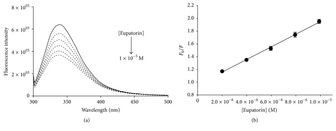
Figure 3.
(a) Fluorescence quenching spectra of reduced PDIA3 alone (solid line) and after stepwise addition of eupatorin (dotted line) (pH 7.4, 25°C, and λ ex = 290 nm). [PDIA3] = 0.5 × 10⁻⁶ M, [eupatorin] = 2 × 10⁻⁵ M final concentration. (b) Stern-Volmer plot of quenching data as mean of at least three independent experiments (standard deviations were better than 10% and correlation coefficient was better than 0.99).

