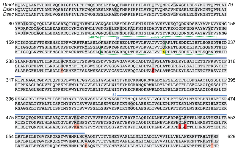
Extended Data Fig. 3. Alignment of drosophilid IR75a orthologues.
Protein sequence alignment of D. melanogaster, D. simulans and D. sechellia IR75a. Blue bars indicate the S1 and S2 lobes of the predicted ligand-binding domain (LBD). The position of the premature termination codon (X) is highlighted in yellow. Dark grey columns in the alignment highlight amino acids conserved only in two of the three species. Pink/red shading represent D. sechellia-specific amino acid changes within the LBD; red are the subset located in the internal cavity of the binding pocket (Fig. 4a). The locations of the peptide epitopes for the IR75a antibodies are highlighted with green dashed boxes.

