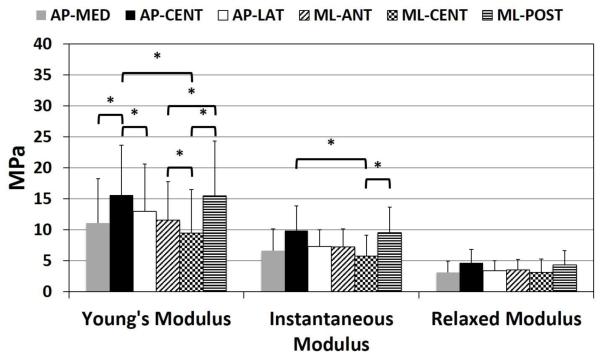
Figure 5.
Young’s Modulus adjusted for strain increment, Instantaneous Modulus and Relaxed Modulus for each region in the AP direction (AP-MED, AP-CENT, AP-LAT) and ML direction (ML-ANT, ML-CENT, ML-POST). Young’s Modulus showed significant differences by region (p<0.001). In the anteroposterior direction, Young’s Modulus was significantly higher in the AP-CENT region compared to both AP-LAT (p=0.04) and AP-MED (p<0.001) regions. In the mediolateral direction, Young’s Modulus, adjusted for strain increment, was significantly lower in the ML-CENT region compared to both ML-POST (p<0.001) and ML-ANT (p=0.04) regions, and ML-ANT was significantly less than ML-POST (p=0.04). In the central region of the articular disc, Young’s Modulus was significantly higher in the anteroposterior direction compared to the mediolateral direction (p=0.04). Differences in Instantaneous Modulus by region were significant (p=0.007). Anteroposteriorly, AP-CENT specimens had a higher Instantaneous Modulus compared to AP-MED specimens, however not significantly (p=0.068). In the mediolateral direction, ML-POST specimens had a significantly higher Instantaneous Modulus compared to ML-CENT specimens (p=0.002). In the central region of the articular disc, Instantaneous Modulus was significantly higher in the anteroposterior direction compared to the mediolateral direction (p=0.001). Differences in Relaxed Modulus by region were not significant (p=0.094).

