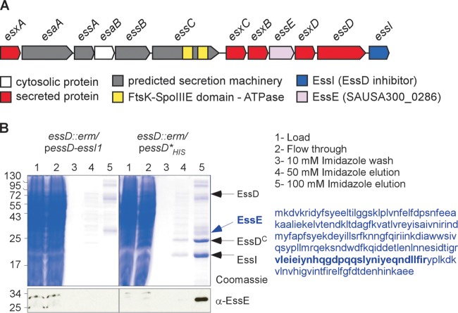
FIG 1.
EssE is a ligand of EssD. (A) Schematic representation of the ESS cluster in S. aureus. (B) Cultures of S. aureus strain USA300 essD::erm carrying plasmid pessD-essI1 or pessD*His to produce wild-type EssD or the nontoxic Leu546Pro variant with a C-terminal histidine tag were grown at 37°C and centrifuged, and sedimented bacteria were lysed to generate cleared lysates that were treated with DDM to solubilize membrane proteins for purification over Ni-NTA (lanes 1). The flowthrough containing unbound proteins (lanes 2), 10 mM imidazole wash (lanes 3), and the 50 and 100 mM imidazole elution fractions (lanes 4 and 5) were separated by SDS-PAGE and either stained with Coomassie blue or transferred to PVDF membrane for immunoblot analyses with the anti-EssE polyclonal serum. Numbers to the left indicate the mobility of molecular mass markers. Arrows point to bands corresponding to proteins identified by mass spectrometry. The sequence of EssE is shown in blue, and the region identified by mass spectrometry is in bold.

