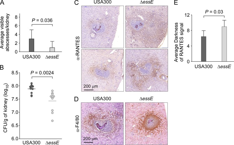
FIG 6.
essE mutants exhibit altered abscesses. Staphylococcal replication in kidneys was measured 5 days postinfection after injection of 5 × 10⁶ CFU of USA300 or its isogenic essE mutant in mice (groups of 8 to 10). (A and B) Kidneys were removed during necropsy to enumerate surface abscesses and subsequently homogenized in 1% Triton X-100 for plating and enumeration of CFU. Statistical analyses were performed using an unpaired two-tailed Student's t test with Welch's correction (A) and a two-tailed Mann-Whitney test (B). (C to E) Kidney sections were taken at 12-μm intervals, and abscesses were first stained with H&E to identify abscesses with staphylococcal communities (dark purple and denser stain in the center of slides) surrounded by immune cells. Next, the sections were incubated with anti-RANTES antibody or F4/80 antibody, a macrophage-specific marker. Following incubation with a secondary HRP-conjugated antibody and development, brown staining reflected the presence of macrophages outside the fibrin cuffs and surrounding USA300 lesions while the staining penetrated ΔessE lesions. Representative images are shown. Anti–RANTES signals were quantified using Fiji software, and values are reported as average darkness. Statistical analysis was performed using a two-tailed Mann-Whitney test.

