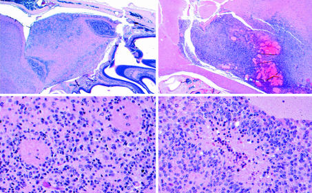
Fig. 2.
Histology of astrocytoma in the resistant NPcis-129 strain compared with the susceptible F1 progeny of NPcis-129 crossed to WT B6. (Left) One of the two GEM WHO III tumors found in NPcis-129 mice (n = 33). The tumor is confined to a focal area of the ventral olfactory bulb (Upper Left). Atypical nuclei diffusely infiltrate the olfactory bulb (Lower Left) and mitotic figures are rare (not shown). (Right) One of three GEM WHO IV tumors found in F1 NPcis-129XB6 progeny (n = 22). This tumor is characterized by infiltrative boundaries (Upper Right), a high mitotic index, and pseudopallisading tumor cells around areas of necrosis (Lower Right).

