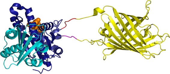
FIG 2.

Structural model of FtsZ with YFP inserted between G55 and Q56. The FtsZ structure is based on PDB file 2VAW (Pseudomonas aeruginosa FtsZ [27]). The N-terminal subdomain is shown in dark blue, the C-terminal subdomain is shown in cyan, and GDP is shown by orange spheres. YFP (PDB entry 1YFP) is shown in yellow. The 3- and 11-aa tails of YFP were added as extended peptides, and the 5-aa linkers are shown in magenta and red. The figure was prepared by the PyMOL Molecular Graphics System, version 1.8 (Schrödinger, LLC).
