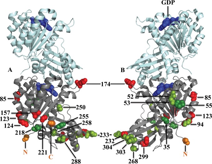
FIG 7.
Cartoon model showing two subunits of an FtsZ pf (PDB entry 3VOA [58]; Staphylococcus aureus FtsZ). The bottom subunit shows sites where the 10-aa GSTLELEGST peptide was inserted. (A) View of the front of FtsZ, from which the C terminus emanates (orange). (B) View of the back of FtsZ, from which the N terminus emanates (orange). Proteins with insertions at dark green sites provided complementation, those with insertions at light green sites generated suppressors, and those with insertions at red sites failed to function in vivo. GDP is shown in blue. The amino acids preceding inserts are shown as spheres, and the E. coli amino acid numbers are indicated.

