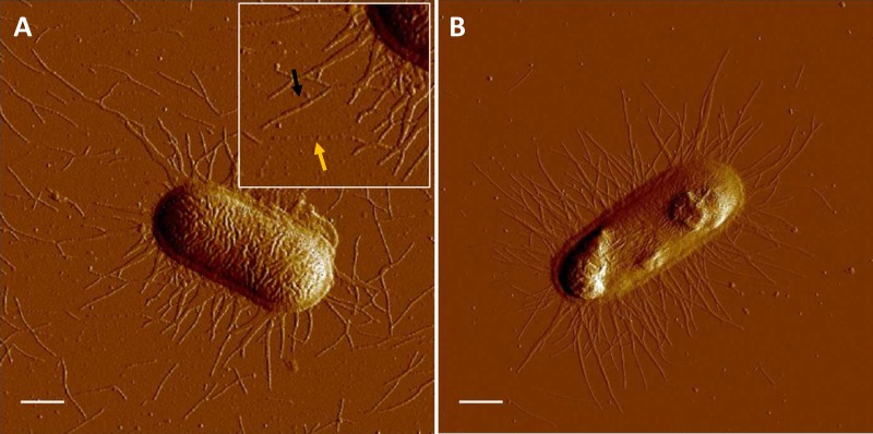
FIG 1.
AFM of ETEC cells expressing fimbriae. (A) Micrograph showing a single C91F cell expressing CS2 fimbriae. The black arrow indicates a fimbria in its helical form (wound), whereas the yellow arrow shows an extended (unwound) CS2 fimbria. The inset represents 1.5× magnification of the micrograph in the vicinity of the arrows. (B) Micrograph showing a single BL21-A2/pMAM2 expressing peritrichous CFA/I fimbriae, which were primarily in their helical form (wound). Bars = 0.5 μm.

