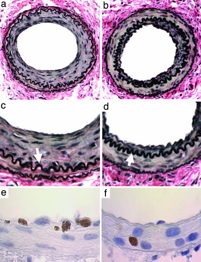
Fig. 4.
Photomicrographs of mouse femoral arteries after injury. (a-d) VerHoeff elastin stain 28 d after injury: (a) wild-type; (b) Nox2-/- (original magnification, ×38); (c) wild-type; (d) Nox2-/- (×150). Neointima separates the internal elastic lamina (arrows) from the lumen. (e and f) Proliferating (BrdUrd-positive) cells 7 d after injury: (e) wild-type; (f) Nox2-/- (×150).

