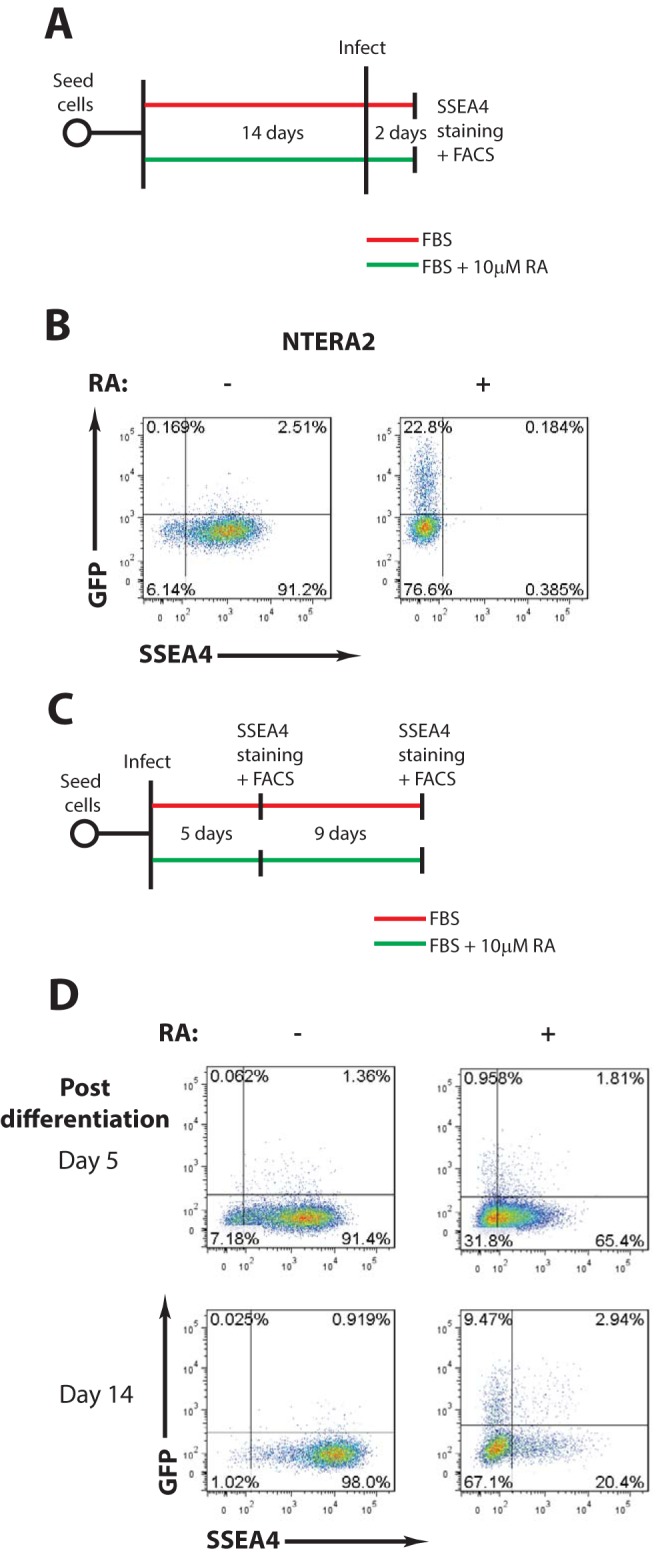
FIG 2.

The block to MLV infection is relieved by differentiation of human EC cells (A) Scheme of experimental setup. (B) Flow cytometry analysis of undifferentiated and retinoic acid-differentiated NTERA2 cells infected with VSV-G-pseudotyped MLV-GFP reporter viruses. Results shown are at day 2 postinfection. The x axis shows SSEA4 staining (a marker of pluripotency); the y axis shows GFP intensity. One representative experiment of three independent experiments is shown. (C) Scheme of experimental setup. (D) NTERA2 cells were infected with VSV-G-pseudotyped MLV-GFP reporter viruses. At 1 day postinfection, cells were cultured with or without retinoic acid to induce differentiation. Flow cytometry analysis was carried out at the indicated times postdifferentiation to monitor MLV gene expression. The x axis shows SSEA4 staining (a marker of pluripotency); the y axis shows GFP intensity. One representative experiment of two independent experiments is shown. RA, retinoic acid; FACS, fluorescence-activated cell sorting.
