FIG 3.
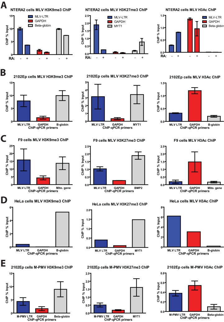
MLV silencing in human EC cells is associated with deposition of repressive chromatin marks. (A) Undifferentiated NTERA2 cells or retinoic acid (RA)-induced differentiated NTERA2 cells infected with MLV-GFP for 3 days were subjected to ChIP analysis using the indicated antibodies. ChIP data are presented as the percentage of input DNA. Results shown are means ± standard deviations (SDs) from two independent experiments performed in duplicate. (B) 2102Ep cells infected with MLV-GFP for 5 days were subjected to ChIP analysis using the indicated antibodies. ChIP data are presented as the percentage of input DNA. Results shown are means ± SDs from three independent experiments performed in duplicate. (C) Experiment similar to that in panel A but performed in F9 mouse EC cells. (D) Experiment similar to that in panel A but performed in HeLa cells. (E) 2102Ep cells infected with M-PMV–GFP for 5 days were subjected to ChIP analysis as described above. qPCR, quantitative PCR.

