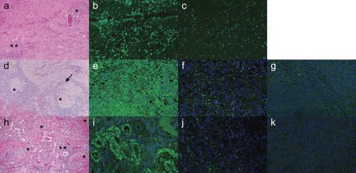
FIG 4.
Paraffin-embedded sections of skin (a to c), lymph node (d to g), and kidney (h to k) tissues stained with H&E (a, d, h) or PCV3 MAb 14 (b, e, i) from porcine dermatitis and nephropathy cases. (a) Sections of skin are characterized by necrotizing vasculitis with fibrinoid changes (*) and scattered lymphoplasmacytic dermatitis (**). (b) The dermal lymphocytic infiltration showed marked intracytoplasmic immunostaining against PCV3. (d) There is a multifocal granulomatous lymphadenitis (*) with the presence of multinucleated giant cells (arrow). (e) The follicular and perifollicular lymphocytic population showed diffuse intracytoplasmic staining against PCV3. (h) Kidneys are characterized by the presence of diffuse membranoproliferative glomerulonephritis (*) with minimal interstitial lymphoplasmacytic infiltration (**). (i) The tubular epithelium showed random areas of positive staining against PCV3. Negative staining of each tissue and background controls were performed by elimination of primary PCV3 MAb 14 and replacing it with PBS, followed by a goat anti-mouse secondary antibody (c, f, j). Lymph node and kidney tissues obtained from animals PCV3 negative by qPCR were concurrently stained with PCV3 MAb 14 and the goat anti-mouse secondary antibody to control for nonspecific antibody binding (g, k).

