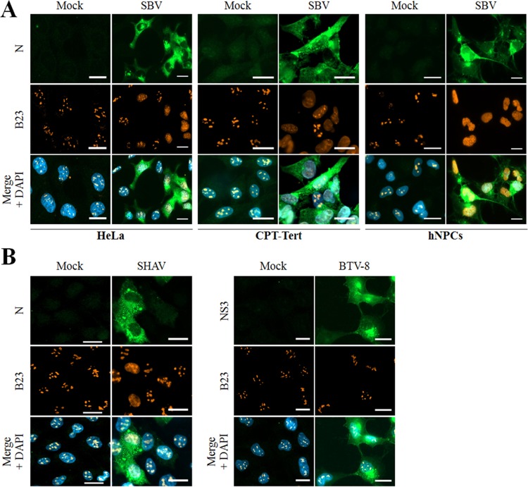
FIG 4.
SBV and a Simbu-related virus trigger nucleolar reorganization during infection. (A) HeLa and CPT-Tert cells and hNPCs were mock infected or infected with the SBV WT (MOI of 0.01). After overnight incubation, cells were fixed with 4% PFA and labeled with DAPI to stain nuclei or stained with specific antibodies for B23 and SBV N. Intracellular localization of DAPI-stained nuclei (blue), B23 (red), and SBV N (green) was visualized by fluorescence microscopy (×63 magnification). Scale bars represent 20 μm. (B) HeLa cells were mock infected or infected with either Shamonda virus (SHAV [left panel]) or bluetongue virus (BTV [right panel]). After overnight incubation, cells were fixed with 4% PFA and labeled with DAPI to stain nuclei or stained with specific antibodies for B23, N SHAV, or NS3 BTV-8. Intracellular localization of DAPI-stained nuclei (blue), B23 (red), and SHAV N or BTV NS3 (green) was visualized by fluorescence microscopy (×63 magnification). Scale bars represent 20 μm.

