FIG 7.
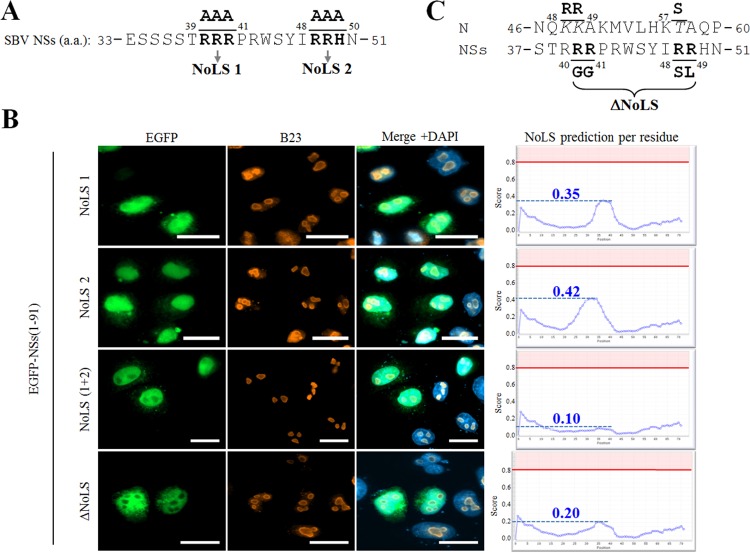
Mutation of a putative nucleolar localization signal impairs NSs nucleolar localization. (A) Amino acid sequence of the aa 33–51 region of SBV NSs. Bipartite triplet of basic residues corresponding to putative NoLS are depicted in bold type and termed NoLS 1 and NoLS 2. (B) HeLa cells were transfected with pEGFP-NSs(1–91) constructs mutated in NoLS 1, NoLS 2, both NoLS 1 and 2, and ΔNoLS as indicated. At 24 h posttransfection, cells were fixed with 4% PFA and labeled with DAPI to stain nuclei or stained with specific antibody for B23. Intracellular localizations of EGFP constructs (green), DAPI-stained nuclei (blue), and B23 (red) were visualized by fluorescence microscopy (×63 magnification). Scale bars represent 20 μm. Each corresponding NoD graphic (right) shows the average NoLS prediction score in the mutated SBV NSs protein. The horizontal red line at value 0.8 of the y axis represents the threshold for NoLS prediction, and the blue dotted line represents the score reached by mutated SBV NSs. (C) Amino acid sequence of the aa 37–51 region of NSs and aa 46–60 region of N. Minimal NoLS mutations designed to be introduced in a recombinant virus were termed ΔNoLS and are depicted in bold type under the NSs sequence. Modified amino acids in NSs sequence appear in bold type, and consecutive changes in N sequence are represented in italic above the NSs sequence.

