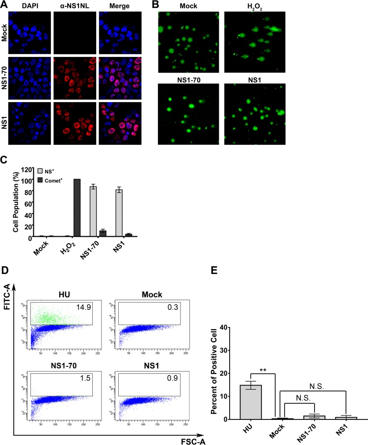
FIG 7.
HBoV1 NS1 and NS1-70 do not induce significant cellular DNA damage. (A to C) Comet assay. HEK293 cells were transduced with HBoV1 NS1- or NS1-70-expressing lentivirus. H2O2 treatment was used as a positive control. At 48 h postransduction, cells were collected for IF analysis and the Comet assay. (A) IF analysis. Half of the cells were stained with anti-HBoV1 NS1NL antibody (red). Nuclei were stained with DAPI (blue). Confocal images were taken at a ×100 magnification. (B) Comet assay. The remaining cells were analyzed for damaged DNA using the Comet assay. Confocal images were taken at a magnification of ×40. (C) Quantification of Comet assay results. Statistical analysis of the percentage of cells that contained damaged DNA and were positive for NS1NL (Comet+) on the basis of the results three independent experiments was performed. Data are shown as means ± standard deviations. (D and E) TUNEL assay. (D) HEK293 cells were mock transduced or transduced with HBoV1 NS1- or NS1-70-expressing lentivirus. At 48 h postransduction, HU-treated cells and the transduced cells were collected and the TUNEL assay was performed, followed by flow cytometry. FITC, fluorescein isothiocyanate; FSC, forward scatter. (E) The percentages of TUNEL-positive cells in each group were quantified and are shown as averages and standard deviations, which were generated from three independent experiments. P values were calculated using Student's t test (**, P < 0.01; N.S., no statistically significant difference [P > 0.1]).

