FIG 8.
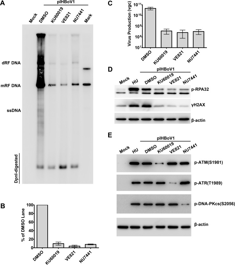
Inhibition of phosphorylation of ATM, ATR, or DNA-PKcs significantly impairs replication of the HBoV1 DNA. HEK293 cells were treated with DMSO or the indicated inhibitor for 4 h prior to transfection with pIHBoV1. At 48 h posttransfection, cells were collected for Southern and Western blot analyses. HU-treated cells were used as a positive control for DDR induction. (A and B) Southern blotting. (A) Representative Southern blot of Hirt extracts probed for HBoV1 NS and Cap. The detected bands are labeled as follows: dRF, double replicative form; mRF, monomer replicative form; and ssDNA, single-stranded DNA. (B) Quantification of mRF DNA in the blot following normalization to the amount of DpnI-digested DNA. The data are based on the results of three independent experiments. Means and standard deviations are shown. (C) Quantification of progeny virus produced. Virus was purified from HEK293 cells transfected with pIHBoV1 for 48 h. Shown are the average numbers of vgc of purified progeny virions per preparation and standard deviations. (D and E) Western blot analysis. (D) Cell lysates were blotted using anti-p-RPA32. The blot was reprobed with anti-γH2AX and β-actin, in that order. (E) Cell lysates were blotted using anti-p-ATM(S1981), anti-p-ATR(T1989), anti-p-DNA-PKcs(S2056), and β-actin.

