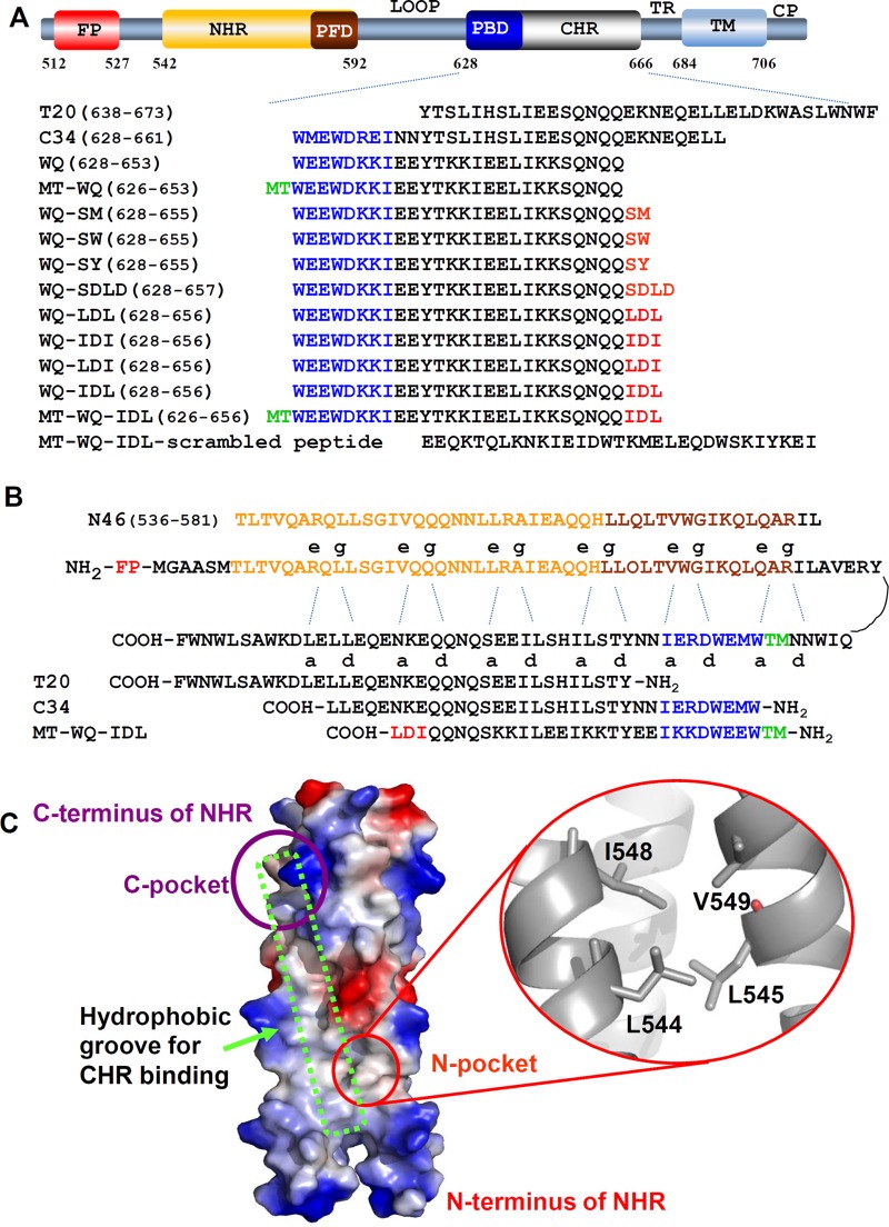
FIG 1.
Schematic illustration of HIV-1 gp41 and 6-HB. (A) Domain distribution of HIV-1 gp41. CP, cytoplasm region; TM, transmembrane region; TR, tryptophan-rich region; CHR, C-terminal heptad repeat; PBD, pocket binding domain; NHR, N-terminal heptad repeat; PFD, pocket-forming domain; FP, fusion peptide region. (B) NHR and CHR bind through residues at the a and d sites and the e and g sites. (C) Crystal structure of gp41 NHR trimer (PDB 2X7R) is shown as an electrostatic surface. Red circle indicates N-terminal hydrophobic pocket formed by aa 544 to 549 of NHR, a potential new binding target on gp41 NHR for a fusion/entry inhibitor. Purple circle indicates the NHR C-terminal hydrophobic pocket. Green box indicates the hydrophobic groove for CHR binding.

