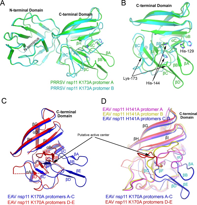
FIG 3.
Differences between nsp11 protomers. (A and B) Structural difference between the two protomers of the PRRSV nsp11 K173A mutant, especially in the CTD. The active sites His129, His144, and Lys173 are shown, and dashed lines indicate residues that have not been resolved in the structures. (C) Two different types of conformations in the CTD of EAV nsp11 K170A mutant protomers. (D) All conformations in the CTDs of EAV nsp11 mutants. Major differences occur in the putative active center.

