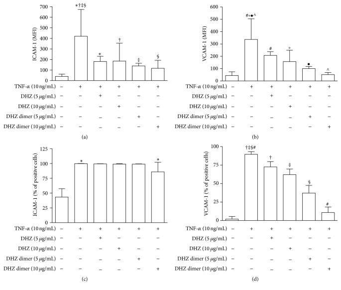
Figure 3.
Cytofluorimetric analysis of adhesion molecule surface expression. HUVEC were pretreated with DHZ or DHZ dimer and then treated with TNF-α 10 ng/mL for 6 hours. (a) and (b) show ICAM-1 and VCAM-1 expression evaluated as mean fluorescence intensity (MFI) (mean values of six experiments); (c) and (d) show ICAM-1 and VCAM-1 expression evaluated as percentages of positive cells (mean values of six experiments). Error bars represent SD. (a) ∗ p = 0.0242; † p = 0.0350; ‡ p = 0.0061; § p = 0.0047. Medium versus TNF-α, p < 0.01. (b) # p = 0.0242; °p = 0.0221; ● p = 0.0061; ∧ p = 0.0012. Medium versus TNF-α, p < 0.001. (c) ∗ p = 0.0061. Medium versus TNF-α, p < 0.001. (d) ‡, # p = 0.0012; †, § p = 0.0061. Medium versus TNF-α, p < 0.001.

