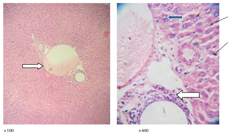
Figure 7.
Photomicrograph of a liver section of mice infected with P. berghei and treated with A. viridis showing no vascular congestion; there is no infiltration of the portal vein; however, the portal veins are mildly inflamed (white arrow). There are scanty inflammatory cells infiltrating the sinusoids (slender arrow); few hepatocytes exhibit vesicular nuclei (blue arrow); others appear normal.

