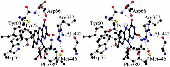
Fig. 4.
Stereo view of the flavin-binding site. The orientation is approximately the same as that of Fig. 3. The flavin has a planar conformation although its N10 atom exhibits a considerable degree of pyramidalization, which positions the C1 atom of the ribityl chain out of the flavin plane. Carbons are shown in black, oxygens are shown in red, and nitrogens are shown in blue. Ordered water molecules are shown as red spheres. H-bond interactions are outlined by the dashed lines.

