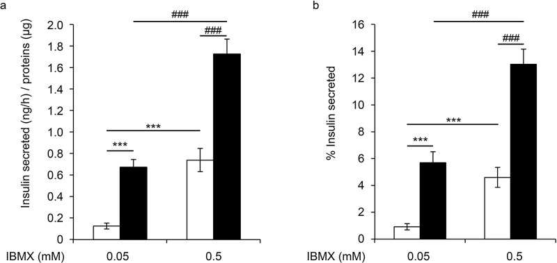
Figure 2.
GSIS on EndoC-βH3 isolated cells and PIs at day 4. Insulin secretion was first stimulated for 60 min with 15 mM glucose in the presence or absence of 3-isobutyl-1-methylxanthine (IBMX; black bars). The medium was then replaced with KRB that contained 2.8 mM glucose (white bars). (a) Data are expressed as secreted insulin in ng per hour/μg of proteins ± SEM of four independent wells per condition assayed in duplicates. (b) Results are expressed as the mean percentage ± SEM of the secreted insulin that was secreted in 1 h of four independent wells per condition assayed in duplicates by ELISA. ***p < 0.001, ###p < 0.001.

