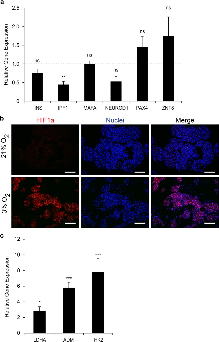
Figure 5.
Molecular impact of hypoxia on the EndoC-βH3 PIs. (a) Expression of β-cell markers in EndoC-βH3 PIs cultured under hypoxia versus normoxia. RT-qPCR was normalized relative to cyclophilin A expression, and the relative expression of each β-cell marker was arbitrarily set to 1 for the control EndoC-βH3 PIs. Each point represents the mean ± SEM of two independent experiments performed in duplicates. **p < 0.01. INS, insulin; IPF1, insulin promotor factor 1 [also known as pancreatic and duodenal homeobox 1 (PDX1)]; MAFA, v-maf musculoaponeurotic fibrosarcoma oncogene homolog A; NEUROD1, neurogenic differentiation factor 1; PAX4, paired box protein 4; ZNT8, zinc transporter 8. (b) Detection by immunohistochemistry of hypoxia-inducible factor 1α (HIF1α) in EndoC-βH3 PIs cultured under hypoxia versus normoxia. Scale bars: 200 μm. (c) Expression of HIF1α target genes was analyzed by RT-qPCR. Each point represents the mean ± SEM of two independent experiments performed in duplicates. *p < 0.05, ***p < 0.001. LDHA, lactate dehydrogenase A; ADM, adenomedullin; HK2, hexokinase 2.

