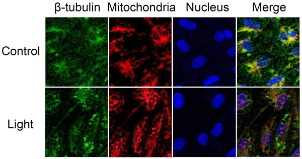
Figure 7.
Cytoskeleton under Stress Conditions. ARPE19 cells were treated under intense light (7,000 lux) for one hr. Expression changes of cytoskeletal proteins, including tubulin, actin, and vimentin, were visualized by immunocytochemistry. Immunocytochemistry using β-tubulin antibody showed colocalization of tubulin with mitochondria. Western blot analysis shows that neurofilament, vimentin, and tubulin changes their expression levels, whereas actin maintains its concentration under oxidative stress (data not shown).

