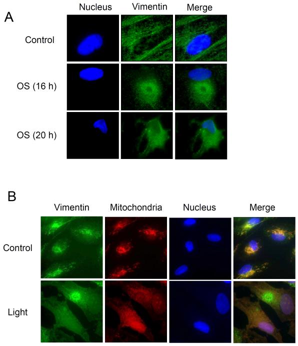
Figure 9.
Intermediate Filaments under Stress Conditions. A. RPE cells were incubated under oxidative stress (tert-BuOOH, 200 μM) for 24 hrs and cells were visualized by immunocytochemistry. B. RPE cells were incubated in the dark (control) or intense light (7,000 lux). Immunocytochemistry using anti-vimentin antibody, Alexa488 secondary antibody, MitoTracker, and DAPI staining showed vimentin (green) and the nucleus (blue).

