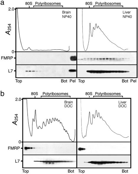
Fig. 1.
Analyses of brain polyribosomes prepared from adult mice. (a) Postmitochondrial fraction was treated with 1% nonionic detergent Nonidet P-40, and total polyribosomes were first concentrated by ultracentrifugation, resuspended, and analyzed by sedimentation velocity throughout sucrose density gradients. Liver polyribosomes treated in the same way were analyzed in parallel. Note the presence of brain polyribosomes and FMRP at the bottom of the centrifuge tube, whereas the distribution of liver polyribosomes is different. (b) Treatment with 1% DOC allows brain and liver polyribosomes to distribute throughout the gradients according to their sedimentation values, whereas FMRP is detected at the top of the gradients. The integrity and distribution of polyribosomes were based on the UV profile as well as the presence of L7, a core protein of the large ribosomal subunit. Distribution of FMRP in different fractions was revealed by immunoblotting with mAb1C3.

