Fig. 3.
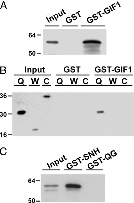
In vitro column-binding assay. (A and B) HA-tagged full-length GRF1 protein (A) or HA-tagged GRF1 fragments (B) were applied to a GST or GST-GIF1 column, as indicated. After extensive washing with the binding buffer to remove nonspecifically bound protein, HA-tagged GRF1 or its fragments retained on the column were eluted and subjected to immunoblot analysis with anti-HA antibody. Q, W, and C above the lanes indicate the GRF1 fragments containing the QLQ, WRC, and C-terminal regions, respectively. (C) The full-length HA-GRF1 protein was applied to a GST-SNH or GST-QG fusion column followed by immunoblot analysis with anti-HA antibody. “Input” indicates the signal elicited by 1% of applied protein as a control.

