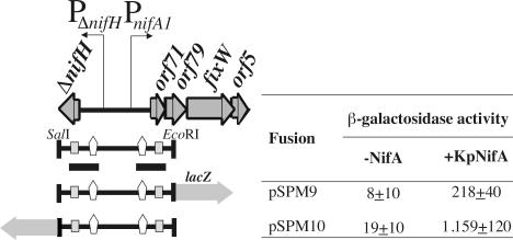
FIG. 6.
NifA-dependent expression of PΔnifH and PnifA1 promoters in E. coli. The physical and genetic map of a 1.3-kb SalI/EcoRI fragment containing the PΔnifH and PnifA1 promoters is shown on the left. The positions of NifA upstream binding sequences (UAS) and σ54-binding sites are indicated by open vertical arrows and grey boxes, respectively. The fragment fused to the lacZ gene in both orientations is shown at the bottom. β-Galactosidase activities associated with the lacZ fusions were measured in E. coli strain ET8000 expressing K. pneumoniae nifA (KpNifA) from plasmid pMJ220. The values (in Miller units) are averages ± standard deviations for three assays.

