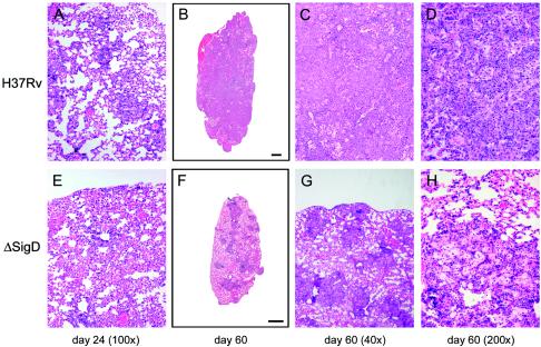
FIG. 9.
Histopathology of lungs of BALB/c mice infected with M. tuberculosis H37Rv or the ΔsigD strain. The images show hematoxylin and eosin staining of lung sections from mice infected with H37Rv (A to D) or the ΔsigD mutant (E to H). The time points at which the mice were sacrificed are indicated below the columns of panels.

