
Figure 5.
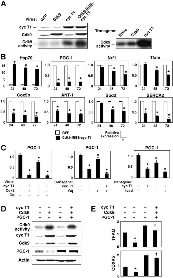
Cyclin T1/Cdk9 suppresses PGC-1, thereby downregulating other genes for mitochondrial function. (A) Left, Western blot and immune complex kinase assays, showing levels of cyclin T1 and Cdk9 48 h after viral gene transfer to cardiac myocytes and their synergistic effect on CTD phosphorylation. Right, Myocardium from wild-type, αMHC-Cdk9 mice, and αMHC-cyclin T1 mice, for comparison. (B) Cyclin T1/Cdk9 suppresses multiple genes for mitochondrial function. RNA from cardiomyocytes 24–72 h after gene transfer was subjected to QRT–PCR analysis. (C) Suppression of PGC-1 mRNA in cultured cardiac myocytes (left) and mouse myocardium (center, right). In vivo, both Gq and mechanical load exacerbate the suppression of PGC-1 by cyclin T1. (D) Cyclin T1/Cdk9 markedly impaired PGC-1 protein expression. Coinfection with PGC-1 virus had no confounding effect on cyclin T1 levels, Cdk9 levels, or Cdk9 kinase activity. (E) Downregulation of genes for mitochondrial function was reversed by supplementing PGC-1. Cells were subjected to virus encoding cyclin T1/Cdk9±virus encoding PGC-1. Gene expression was normalized to GAPDH (Handschin et al, 2003). (B, C (left), E) *P<0.05 versus GFP; †P<0.05 versus cyclin T1/Cdk9. (C (right, middle)) *P<0.05 versus nontransgenic littermates.