
Figure 5.
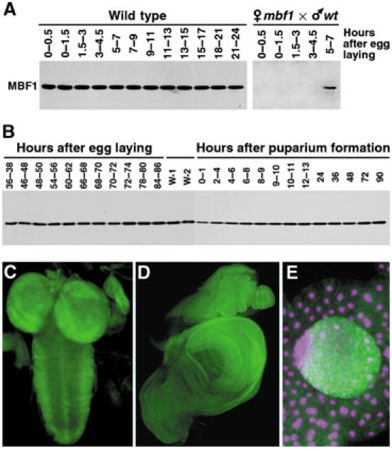
Expression pattern of MBF1 in Drosophila. (A) Western blot showing constitutive MBF1 expression during embryogenesis. Embryos from mbf1 mutant mothers were devoid of all maternal MBF1 protein. Zygotic expression from the paternal wild-type chromosome began between 5 and 7 h after egg laying (right panel). (B) Western blot showing MBF1 expression throughout the postembryonic life. W-1 and W-2, wandering stages of larvae. (C–E) Sites of high MBF1 expression during larval life were the central nervous system (C, second instar), imaginal discs (D, late third instar) and the testis (E, center), but not the fat body (E, surrounding tissue). MBF1 was detected with a specific polyclonal antibody, and DAPI was used for DNA staining in (E). Magenta is used as colorblind friendly (http://jfly.iam.u-tokyo.ac.jp/color/text.html).