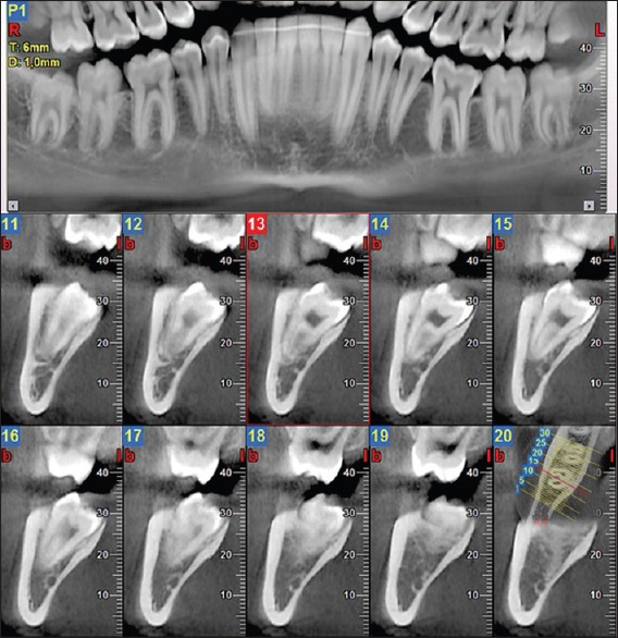
Figure 1.

Cone-beam computed tomography: 1 mm mesiodistally spaced cross-sections at the level of the lower right third molar which is vertically positioned. The third molar roots overlap the inferior alveolar nerve and the canal cortical bone does not show any discontinuity (sections 13 and 14)
