
Figure 9.
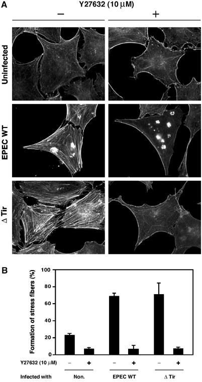
Effect of Y27632 on EPEC-induced actin stress fiber formation. (A) Swiss 3T3 cells were maintained in the presence or absence of 10 μM Y27632 for 1 h, and were infected with EPEC WT and the tir mutant for 3 h in the presence or absence of 10 μM Y27632. The cells were fixed and stained with rhodamine-labeled phalloidin in order to detect F-actin. All fluorescence images were taken with the same exposure time. (B) The percentage of Swiss 3T3 cells showing marked formation of actin stress fibers is indicated. Swiss 3T3 cells were infected with EPEC WT and the tir mutant for 3 h in the presence or absence of 10 μM Y27632 as in (A). Percentages are based on counts of at least 150 cells, and the values are the means±standard deviation from three independent experiments.