
Figure 1.
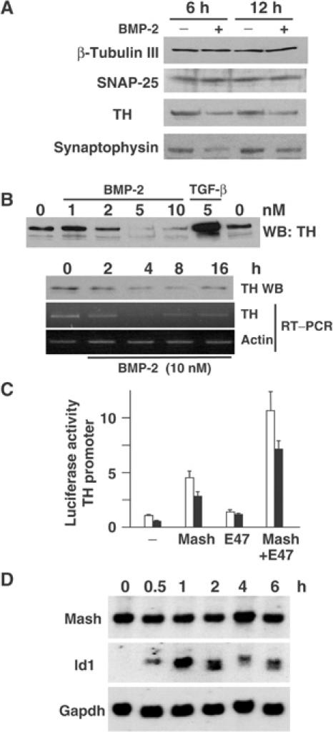
(A) DMS53 cells were treated with 5 nM BMP-2 for 6 or 12 h. Total extracts were obtained and analyzed by immunoblotting for the neural markers indicated. (B) DMS53 cells were treated for 4 h with the indicated amounts of BMP-2 or TGF-β (upper panel) or with 10 nM BMP-2 for the times indicated (lower panel). TH and actin expressions were analyzed by immunoblotting and/or semiquantitative RT–PCR. (C) C17.2 cells were cotransfected with a TH promoter-driven reporter construct and the combinations of Mash1 and/or E47 expression vectors indicated. Luciferase assay was performed after treatment for 16 h with 5 nM BMP-2 (filled bars) or no addition (empty bars). The results are expressed as mean±s.e.m. of triplicates from four independent transfections. (D) Northern blot analysis was performed with total RNA from DMS53 cells treated for the indicated times with 10 nM BMP-2 as described in Materials and methods.