
Figure 3.
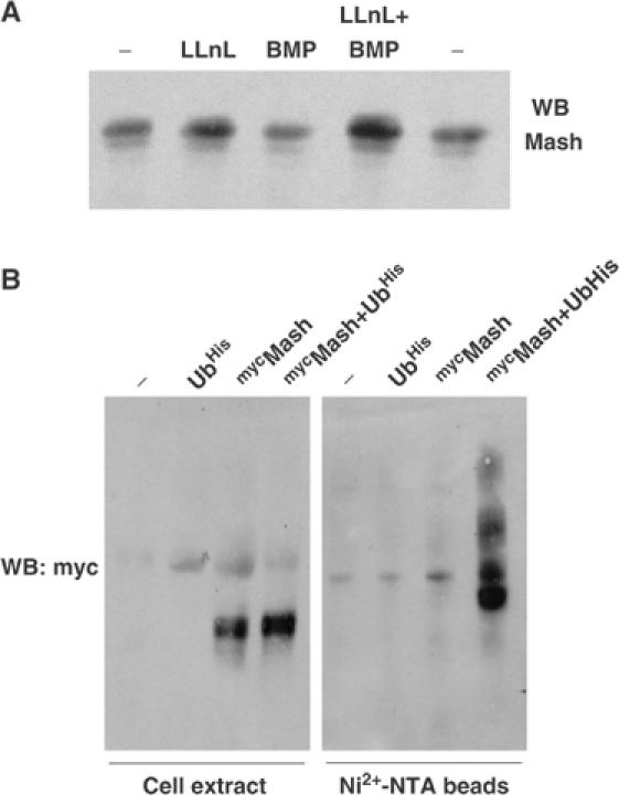
(A) DMS53 cells were treated with 50 μM LLnL and/or 5 nM BMP-2 for 2 h and expression of Mash1 was analyzed by immunoblotting. (B) Extracts from HEK-293 cells transfected with the indicated expression constructs and treated overnight with 50 μM LLnL were subjected to immunoblotting to detect myc-Mash1 expression before (left panel) or after Ni2+-NTA-agarose purification (right panel).