
Figure 4.
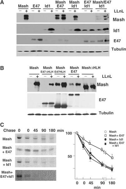
(A, B) Extracts from HEK-293 cells transfected with the expression constructs indicated, treated or not with 50 μM LLnL for 8 h, were analyzed by immunoblotting. (C) HEK-293 cells, transfected with the constructs indicated, were pulsed with [35S]methionine for 3 h and labeled proteins were chased in unlabeled media for different times. Labeled Mash1 was immunoprecipitated and visualized by SDS–PAGE. Quantification of Mash1 levels from three to four separate assays is expressed as mean±s.e.m. of percentage values of samples at time zero.