
Figure 2.
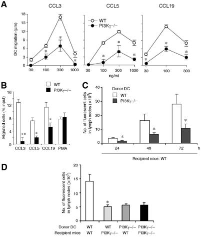
Defective in vitro and in vivo migration of CD34-derived DC from PI3Kγ−/− mice. (A) Migration of iDC (to CCL3, CCL5) and TNF-mDC (to CCL19) was evaluated by the leading-front techniques, using nitrocellulose filters. The results are reported after subtraction of basal migration (against medium; 4±2 and 3±2 μm for WT and PI3Kγ−/− DC, respectively). (B) Migration of iDC and mDC was evaluated by Transwell insert. The results are at the net of basal migration: 5±2 and 3±2% for WT and PI3Kγ−/−, respectively. (C) TNF-mDC from WT and PI3Kγ−/− mice were labelled with the vital dye CFSE and injected subcutaneously in the hind leg footpad of WT mice. Popliteal lymph nodes were recovered 24, 48 and 72 h later and the cell suspension was evaluated by flow cytometry. (D) WT and PI3Kγ−/− TNF-mDC were injected either in WT or PI3Kγ−/− mice and recovered after 48 h. In panels C and D, results are expressed as the number of fluorescent cells recovered from the draining lymph nodes. Each experiment is representative of four experiments (**P<0.01, *P<0.05 versus respective WT control group).