
Figure 3.
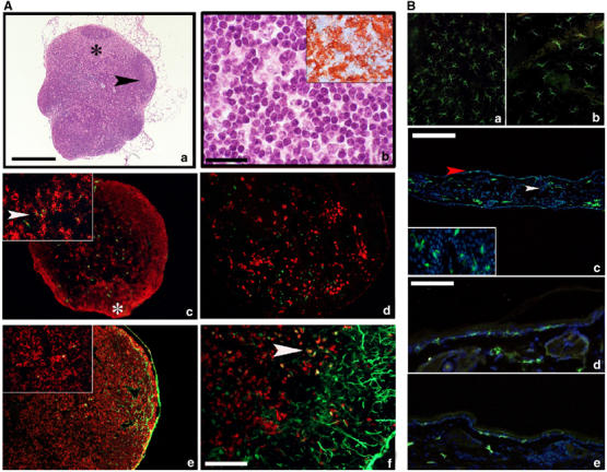
Morphology and DC distribution in PI3Kγ−/− lymph nodes and skin sections under steady-state and inflammatory conditions. (A) Haematoxylin/eosin staining of a lymph node section from PI3Kγ−/− mice (a) displays normal architecture including cortex, with full-blown germinal centre (black arrow), and an expanded paracortex (black asterisk) populated by cells with dendritic morphology (b) with strong expression of MHC-class II (inset in b). Sections from a popliteal lymph node obtained after injection of CFSE-labelled DC immunostained for MHC-class II (red in c). A low-power field reveals two cortical nodules of MHC-class II+ cells representing B-cell follicles (white asterisk) and sparse MHC-class II+ cells with DC morphology, some of which are labelled by CFSE green fluorescent material in their cytoplasm (inset in c, arrow). Langerin-positive cells populate the subcortical area but do not contain CFSE green fluorescent material (d). Sections from an inguinal lymph node obtained after FITC-based skin painting were counterstained with MHC-class II molecule (e) and Langerin (f). The marginal sinus is filled with green fluorescent material and scattered cells in the paracortex contain green dots in their cytoplasm; these cells express MHC-class II+ (inset in e) and some of them also Langerin (f; yellow cells, see arrows). Magnification × 40 (a, c, d and e; scale bar in a: 500 μm), × 100 (f; scale bar: 200 μm), × 200 (b; scale bar: 100 μm) and × 400 (insets). Anti-MHC-class II-positive cells were detected by the immunoperoxidase technique (inset in b) and immunofluorescence (c, e (red staining)); Langerin was detected by immunofluorescence (d, f (red staining)). (B) (a, b) Two representative examples of epidermal sheets respectively from WT and PI3Kγ−/− animals, immunostained for MHC-class II molecule. MHC-class II+ cells show a typical stellate morphology and regular distribution; an evident reduction in number is observed in PI3Kγ−/− epidermis (b). A low-power field from an entire representative section obtained from WT mice ears (c) lined by two thin layers of epidermis. MHC-class II+ green cells are present in the epidermis (red arrow) as well as in the superficial dermis (white arrow) corresponding respectively to LC and to dermal DC. MHC-class II+ LC display fine and long dendrites and multiple interactions with the surrounding keratinocytes (inset). A representative high-power field from WT skin section (d) shows numerous MHC-class II+ LC and dermal DC homogeneously distributed. In PI3Kγ−/− (e), a reduced population of MHC-class II+ LC is observed. Immunofluorescence analyses were performed with FITC-conjugated anti-MHC-class II (green) and nuclei were counterstained with DAPI. Magnification × 40 (c; scale bar: 500 μm), × 200 (a, b, d, e; scale bar in d: 100 μm) and × 400 (inset in c).