
Figure 6.
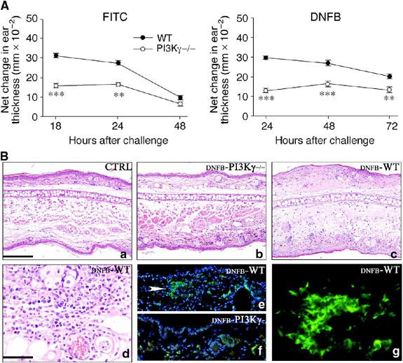
Defective CHS in PI3Kγ−/− mice. (A) WT and PI3Kγ−/− were sensitized with FITC (left panel) or DNFB (right panel) on their shaved abdominal skin on day 0. After 5 days, mice were challenged on the right ear. The left ear was painted with vehicle as control. Increases in ear swelling were measured at different time points and are presented as increased swelling at the net of contralateral ear value. Mean±s.d. for each group (eight mice) are presented (**P<0.01; ***P<0.001 versus respective WT group, by ANOVA Tukey's multiple comparison test). Each experiment is representative of three experiments. (B) Skin sections are obtained from unstimulated control (CTRL) animals (a), and after CHS induction with DNFB (24 h after challenging) respectively from PI3Kγ−/− (DNFB-PI3Kγ−/−) (b, f) and WT (DNFB-WT) (c, d, e, g). Upon CHS induction, only scattered leucocytes infiltrate the dermis of PI3Kγ−/− (b); in contrast, in WT mice, a diffuse dermal oedema is observed in association with microvascular dilatation and dense inflammatory infiltrate; the latter predominantly includes granulocytes, with occasional eosinophils and mononuclear cells (c, d); immunofluorescence demonstrates that the mononuclear cells exhibit a dendritic morphology, form aggregates and express MHC-class II antigens (e, white arrow head and g). This DC population is scant in PI3Kγ−/− and is represented by scattered cells (f). Magnification × 100 (a–c, e, f; scale bar in a: 200 μm) and × 400 (d, g; scale bar in d: 50 μm).