
Figure 3.
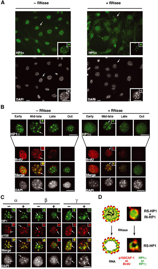
Replication-specific RNase-resistant HP1α and HP1γ but not HP1β pools around pericentric loci colocalizes with p150CAF-1 and BrdU. (A) A subset of cells retain some HP1α staining at pericentric heterochromatin domains following RNase treatment. A wide field showing HP1α labeling (green, top) and DAPI staining (bottom) in 3T3 cells without or with RNase treatment before fixation. The insets show higher magnifications focused on the cell indicated by the arrow as well as on individual foci magnified four-fold. Scale bar, 10 μm. (B) Only cells in mid-late S-phase retain significant HP1α staining as a rim around pericentromeric heterochromatin and sites of DNA synthesis. Double labeling in 3T3 cells of HP1α (green, top isolated) and BrdU (red) in 3T3 cells treated or not with RNase is shown for the early, mid-late, late, and out of S-phase. The arrow indicates typical foci magnified four-fold in the inset. The merge and corresponding DAPI staining are presented. Scale bar as in (A). (C) In mid-late S-phase cells, a fraction of HP1α and HP1γ but not HP1β is RNase-resistant and forms a rim around pericentromeric heterochromatin colocalizing with p150CAF-1. Double labeling of mid-late S-phase 3T3 cells with the three HP1 isoforms (green) and p150CAF-1 (red) without (−) or with (+) RNase treatment. Scale bar and arrows as in (B). (D) Model for HP1α and HP1γ distribution during replication of pericentromeric heterochromatin in mid-late S-phase. Top: Replicating pericentric heterochromatin is represented with a replication-independent pool of HP1 (RI-HP1, large light green circle) in the inner region interacting with RNA (black), and a replication-specific pool of HP1 at the periphery (RS-HP1, small dashed green circles). The location of p150CAF-1 or BrdU incorporation is shown (red circle). Colocalization of the DNA synthesis/p150CAF-1 site and RS-HP1 is represented in yellow. Bottom: Upon RNase treatment, the replication-independent pool of HP1 (RI-HP1) in the inner part is removed following RNA degradation without affecting the replication-specific pool of HP1 (RS-HP1) at the periphery. Corresponding stainings for p150CAF-1 (red) and HP1α (green) are shown as magnified images.