
Figure 4.
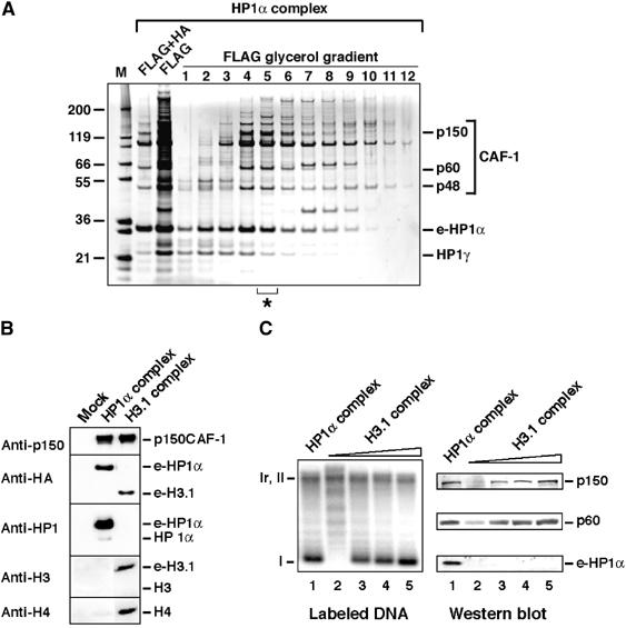
CAF-1 is found in HP1α/γ- and H3.1-containing complexes. (A) Analysis of e-HP1α-containing complexes. SDS–PAGE with fractions purified by FLAG and HA affinity (FLAG+HA), FLAG affinity (FLAG), and from glycerol gradient with the FLAG affinity-purified complex (FLAG glycerol gradient) stained with silver. The asterisk identifies the fraction used in (C). Positions corresponding to CAF-1 p150, p60, p48, HP1γ, and e-HP1α identified by mass spectrometry are indicated. Molecular weight markers (M) are shown. (B) Histones are not detected in the HP1α–CAF-1 complex. Western blot of complexes purified from mock, or e-HP1α- or e-H3.1-transduced HeLa cells. Proteins identified (right) and antibodies used (left) are indicated. (C) Comparison of the HP1α and H3.1 complexes in promoting nucleosome assembly coupled to DNA synthesis. Left: Supercoiling analysis. Cytosolic extract deficient in CAF-1 activity (p150) is used in combination with the HP1α–CAF-1 complex (fraction 5 of the glycerol gradient from (A), lane 1) and increasing amounts of the H3.1–CAF-1 complex (lanes 2–5). The migration of relaxed/nicked (Ir/II) and supercoiled circular DNA (I) is indicated. Right: Western blot analysis with samples corresponding to reactions on the left. Revelation with antibodies is as indicated.