
Figure 5.
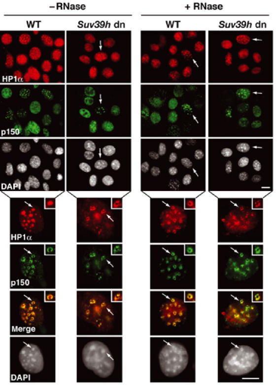
In Suv39h double mutant cells, a replication-specific pool of HP1α is colocalizing with p150CAF-1 at pericentric heterochromatin domains. Top: A wide field showing HP1α labeling (green), and p150CAF-1 (red) and DAPI staining in WT or Suv39h double-null (Suv39h dn) MEFs treated or not by RNase before fixation. The arrows indicate cells in mid-late S-phase based on p150CAF-1 staining. Scale bar, 10 μm. Bottom: As above with a mid-late S-phase nucleus. Merge and DAPI staining are presented. The arrow indicates typical foci magnified three-fold in the inset. Scale bar, 10 μm.