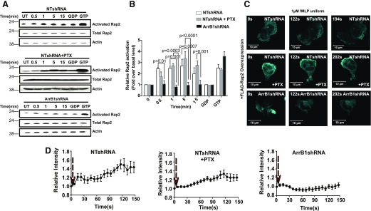
Figure 5. Rap2 activation is independent of Gαi but is dependent on ArrB1.
(A) NTshRNA and NTshRNA-infected cells were pretreated with PTX and ArrB1shRNA stimulated with 1 µM fMLP for the indicated periods of time. Activated Rap2 (Rap2-GTP) was isolated by association with Ral-GDS-RBD immobilized on agarose beads, and captured proteins were detected by Western blots using anti-Rap2-specific antibodies (n = 3). (B) The resulting Western blots were quantified and normalized to Rap2. (C) Activated Rap2 translocated to plasma membrane under fMLP bath stimulation (1 µM) in an ArrB1-dependent manner. Panels show extracted frames from time-lapse confocal microscopy revealing the translocation of eGFP-Ral-GDS from the cytosol to the plasma membrane lamellar edge (7 s intervals). (D) The relative membrane translocation of eGFP-Ral-GDS upon fMLP stimulation in NTshRNA-treated (top; n = 6), NTshRNA + PTX-treated (middle; n = 4), or ArrB1shRNA-treated (bottom; n = 5), differentiated cells.

