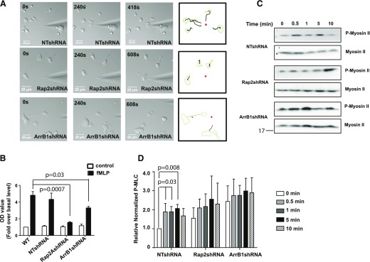
Figure 7. Rap2 GTPase required for neutrophil adhesion.
(A) Rap2- and ArrB1-depleted cells showed defected migration in response to the chemotactic gradient shown in the middle and bottom panels, respectively, when compared with NTshRNA-infected cells (top). Tracings (right) show the migratory paths traveled by cells, as revealed by time-lapse microscopy. (B) Both Rap2- and ArrB1-depleted cells exhibited less fMLP-stimulated adhesion then their uninfected and NTshRNA-infected counterparts. Values are presented as fold over basal level in uninfected cell (n = 3). (C) Rap2- and ArrB1-depleted cells showed defective fMLP-stimulated myosin II activation by using p-MLC-specific antibodies on lysates by Western blot analysis. (D) Quantification revealed no significant stimulation of p-MLC II in Rap2- and ArrB1-depleted cells compared with NTshRNA when normalized to total myosin II on parallel blots. Asterisks indicate needle positions.

