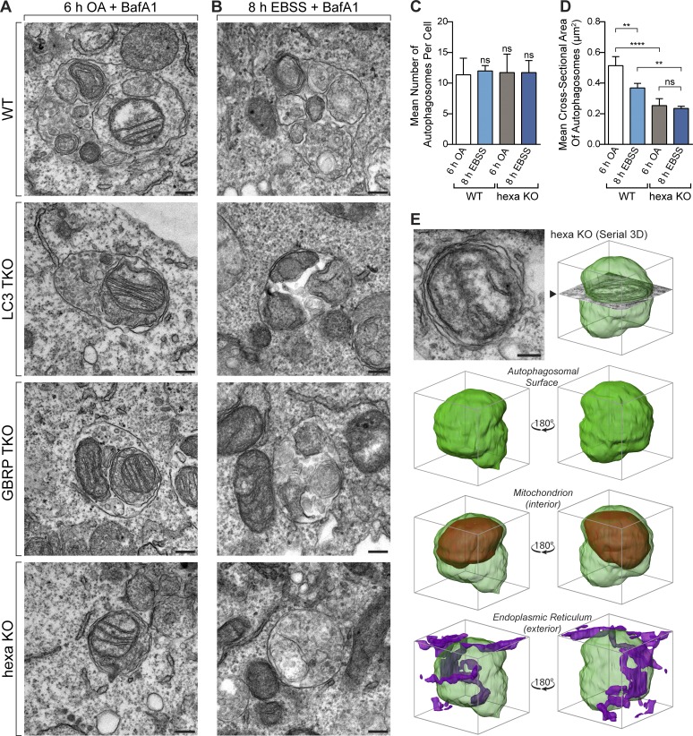
Figure 3.
Atg8s are dispensable for autophagosome formation but regulate autophagosome expansion. (A and B) Representative TEM images of autophagosomes containing mitochondria in WT, LC3 TKO, GBRP TKO, and hexa KO cells after incubation for 6 h with OA and BafA1 (A) or for 8 h with EBSS and BafA1 (B). Wider field-of-view images and untreated examples shown in Fig. S3 D. (C and D) TEM quantification of mean autophagosome number per cell (C) and the mean cross-sectional area of autophagosomes in WT and hexa KO cells treated with BafA1 and either OA for 6 h or starved with EBSS for 8 h (D). Scatterplot of measurements provided in Fig. S4 A. (E) 3D rendering of a reconstructed autophagosomal compartment (green) from a hexa KO cell after 6 h OA and BafA1 treatment, shown with sequestered mitochondrion (red) and endoplasmic reticulum (purple). (Source images and further examples shown in Fig. S3, A–C.) Data in C and D are mean ± SD from three independent double-blinded experiments, using measurements from exactly 12 randomly chosen cells (>100 autophagosomes) per sample. **, P < 0.005; ****, P < 0.0001 (one-way ANOVA). Bars, 200 nm.

