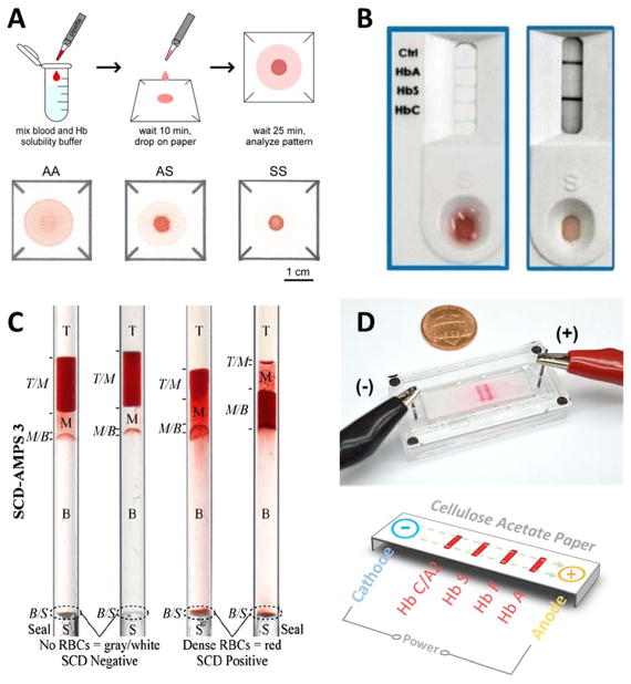
Figure 2. Illustrations of the principle of operation of the emerging technologies for SCD diagnosis.
(A) Paper-based Hemoglobin solubility180. A droplet of blood mixed with Hb solubility buffer is dropped on chromatography paper, and a blood stain is allowed to form. The stain on paper is analyzed and the color intensity profiles are used to determine the Hb type in the sample. (B) Sickle Scan™ lateral flow immunoassay181. The test specimen consisting of a drop of blood mixed with Hb solubility buffer is dropped onto the sample loading zone. The solution then diffuses to the test zones where Hb is captured by color-conjugated antibodies. The type of Hb is determined by the appearance of a blue line at the different test zones along the test strip. (C) Density-based separation184. The blood sample is mixed with aqueous polymeric solutions in capillary tubes. Upon centrifugation, the precipitation of a dense RBC layer at the bottom of the tube indicates SCD. (D) Microengineered electrophoresis (HemeChip). After loading the blood sample mixed with DI water into the chip, an applied electric field causes Hb separation. Due to the differences in mobility among Hb types, each type will travel a unique distance across the paper strip.

