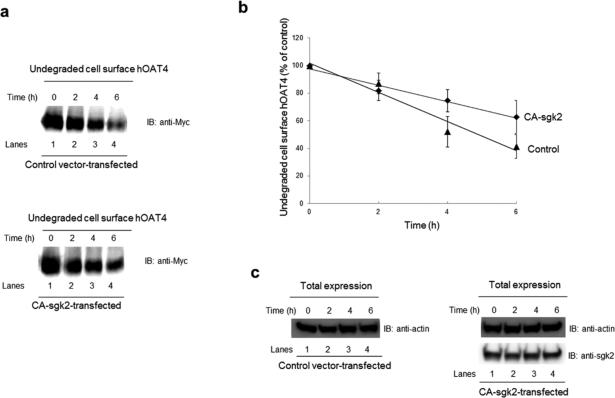
Fig. 4. Effect of sgk2 on the degradation of cell surface hOAT4.
(a) COS-7 cells were cotransfected with hOAT4 and control vector (top panel) or with hOAT4 and the constitutive active form of sgk2 (CA-sgk2) (bottom panel). Cell surface hOAT4 degradation was then analyzed as described in “Materials and Methods” section followed by immunoblotting (IB) using anti-myc antibody. (b) Densitometry plot of results from Fig. 4a as well as from other experiments. The amount of undegraded cell surface hOAT4 was expressed as % of total initial cell surface hOAT4 pool. Values are mean ± S.E. (n = 3). (c). Total expression of sgk2 and house-keeping protein β-actin. COS-7 cells were co-transfected with hOAT4 and control vector or with hOAT4 and the constitutive active form of sgk2 (CA-sgk2). Transfected cells were lysed, followed by immunoblotting (IB) with anti-sgk2 antibody or anti-actin antibody.

