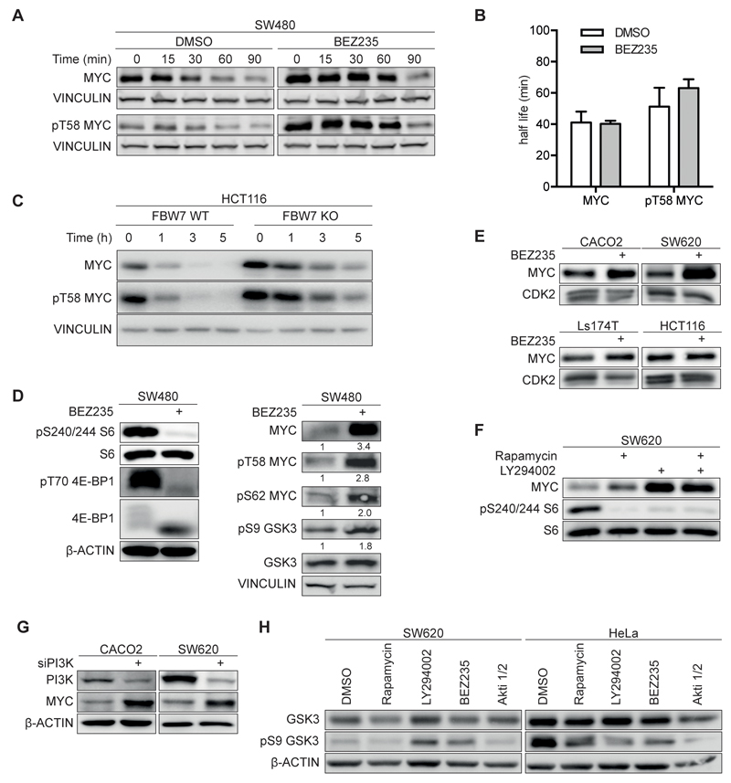
Figure 1. Effect of PI3K/mTORC inhibition on MYC expression and stability in colorectal cancer cells.
A. Immunoblots documenting MYC and pT58 MYC stability. SW480 cells were treated with 200nM BEZ235 or solvent control for 24h and cycloheximide (50µg/ml) and harvested at the indicated time points. Vinculin was used as loading control. Exposures of MYC and pT58 MYC blots were adjusted to equalize exposure at 0 min (n=3; unless otherwise indicated, n indicates the number of independent biological repeat experiments in the following legends).
B. Calculated half-life of total MYC and pT58 MYC. Immunoblots shown in panel A.
C. Immunoblots show MYC and pT58 MYC stability in wild type (WT) and FBXW7 deficient (KO) HCT116 cells (n=1).
D. SW480 cells were incubated with 200nM BEZ235 for 24h. Left panel document effect on mTOR targets S6 and 4E-BP1, right panel on MYC and GSK3 (n=2).
E. Immunoblots of four colorectal cell lines upon treatment with BEZ235 (500nM; 24h) or solvent control (n=3).
F. SW620 cells were treated for 24h with Rapamycin (100nM), LY294002 (50µM) or both and analyzed by immunoblotting.
G. The indicated cell lines were transfected with siRNA targeting the p110alpha subunit of PI3K or control siRNA. 72h after transfection protein levels were determined by immunoblotting (n=2).
H. Immunoblot of cells treated for 24h with indicated inhibitors or solvent control. (Rapamycin 100nM, LY294002 50µM, BEZ235 500nM, Akti 1/2 1µM).

