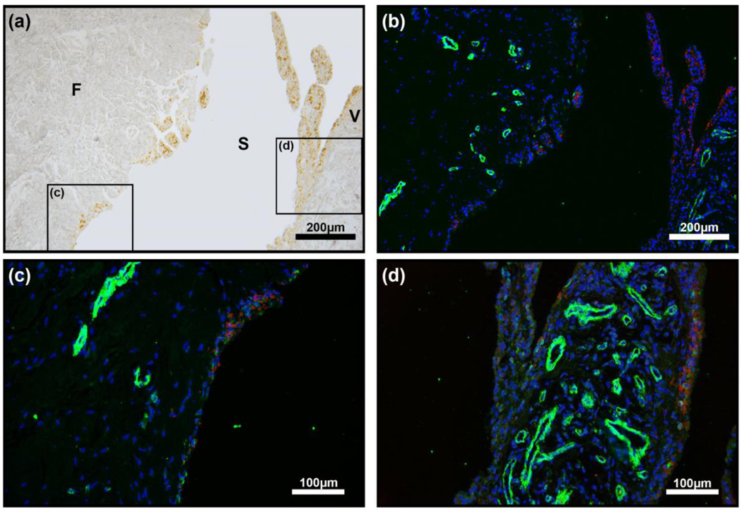
Figure 2.
a-Smooth muscle actin (a-SMA) IHC of synovial tissue in haemophilia patient. (a) Bright field image of synovial tissue showing brown HMCs in the surface layer of synovial membrane. (b–d) a-SMA (green) which is a marker of active myofibroblasts was not readily seen in the synovial tissue of haemophilia patients except surrounding blood vessels in both the fibrous sub-surface layer (F) and synovial villi (V). a-SMA: green; DAPI:blue; HMCs: red.

